AESTHETIC DENTISTRY
Removable prostheses and aesthetics Once teeth are extracted and disposed of, important information is lost for future replacement. The size, shape and surface detail provide data of the natural dentition. It has been advocated that dental practitioners wash extracted teeth, pack them discreetly in a suitable container and offer them to the patient to keep for reference informs Besford and Sutton, 2018. Clinical implication: Ask patients to bring several photographs to the first appointment showing natural teeth. A photometric method is described for selecting front denture teeth: a. Measure the distance between the patient’s pupils (PD). Line up the zero edge of a metal ruler with the right edge of the patient’s right iris. Then, read off the right edge of the left iris in mm. A PD of 65 ± is typical. b. Scan the photograph. Enlarge the image on a screen. Use a ruler or digital capilers to measure the PD on the photography and the width of the upper two central incisors on the photograph. True width = photographic width X true PD Photographic PD A typical result would be 17.6 for the central and lateral incisors. Divide this by two to give the width of a single central incisor. c. Use a mould chart to locate central incisors of approximately the correct width. Practical application: Patients may have dentures whose appearance they really liked and wish to have repeated. There may be a relative who has similar teeth. Dental radiographs taken before the front teeth were extracted may indicate size and shape. Women’s teeth are on average 2% smaller than men’s teeth. Regarding colour selection in the older patient, it is up to the clinician to mention that lighter teeth for an older patient as usually difficult to reconcile as natural. Reference: Besford, J.N. and Sutton, A.F., 2018. Aesthetic possibilities in removable prosthodontics. Part 3: Photometric tooth selection, tooth setting, try-in, fitting, reviewing and trouble-shooting. British Dental Journal 224(7), p.491.
PROSTHODONTICS

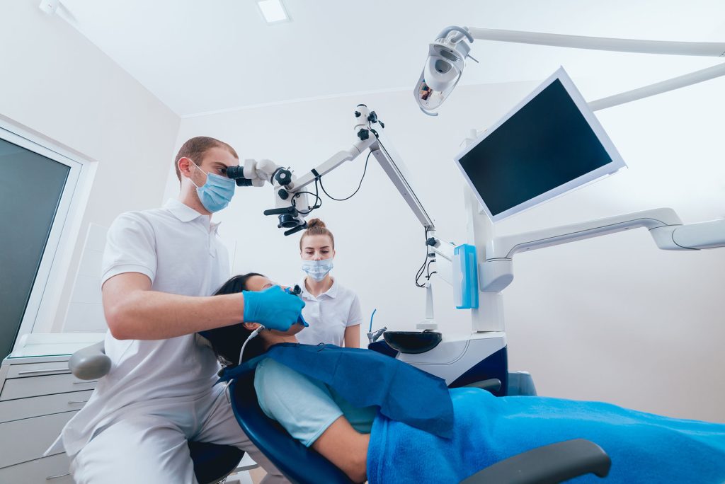
Trigeminal neuralgia revisited Trigeminal neuralgia (TN) is characterised by touch-evoked unilateral brief shock-like paroxysmal pain in one or more divisions of the trigeminal nerve. In addition to the paroxysmal pain, some patients also have continuous pain. A neurovascular conflict which involves morphological changes of the trigeminal nerve is highly associated with classical TN and is present in about half of TN patients. TN can be divided into classical TN and secondary TN. Secondary TN is caused by either multiple sclerosis or a space-occupying lesion affecting the trigeminal nerve reports Maarbjerg et al., 2017. Clinical implication: The diagnosis is based on patient history. 1. Pain onset is important: 1a. If the pain was preceeded by or coincided with a herpes zoster in the ipsilateral trigeminal distribution, painful trigeminal neuropathy attributed to acute herpes zoster should be considered. 1b. If the pain was preceeded by a relevant trauma to the ipsilateral side of the face, e.g. such as invasive dental procedures or fractures, painful post-traumatic trigeminal neuropathy is more likely. 2. Pain location is important: 2a. In bilateral constant pain located in the temporomandibular area, tension-type headache, TMD and persistent idiopathic facial pain should be considered. 2b. If short-lasting intense stabbing pain is isolated to the scalp or occipital area, diagnoses such as occipital neuralgia, primary stabbing headache and paroxysmal hemicranias should be considered. Practical application: An early work-up should include and MRI of the brain and brainstem, ECG and laboratory testing. Generally, first line treatment is prophylactic medication with sodium channel blockers. In medically refractory patients, surgical treatment is the next step. Reference: Maarbjerg, S., Di Stefano, G., Bendtsen, L. and Cruccu, G., 2017. Trigeminal neuralgia–diagnosis and treatment. Cephalalgia, 37(7), pp.648-657.
ORAL MEDICINE

Understanding sickle cell disease in clinical practice Sickle cell disease is one of the most common autosomal recessive genetic diseases associated with production of abnormal haemoglobin. The main clinical features are haemolytic anaemia and vascular occlusion. Chekroun et al., 2019 points out that acute complications are frequent and variable and include chest syndrome, stroke, infection mainly due to abnormal spleen function and bone pain. Oral lesions are common. Clinical application: Oral lesions include aseptic pulp necrosis, mucosal damage due to anaemia, fungal infections due to numerous antibiotic therapies, dental eruption delays, bone pain and oral neuropathies including the mental nerve of the chin caused by infarction of the vascularisation of the lower mental nerve or its branches. Bone lesions may also be visible radiologically throughout the skeleton and at the level of the maxilla. Altered pallor of the oral mucosa is the most frequent oral manifestation observed. Atrophy of the tongue is often seen. Practical implication: Adult sickle cell patients should be seen annually. Mild sickle cell disease does not require particular management. Severe or moderate sickle cell disease requires precautions for dental management: 1. Antibiotic prophylaxis for invasive treatment is needed (endodontics, oral surgery, periodontal surgery, root surface scaling). 2. Clinical and radiological examination to detect oral infectious foci 3. Steroidal anti-inflammatory drugs are contra-indicated. 4. Deep local anaesthesia is suggested and the amount of vasoconstrictor is reduced. Use nitrous oxide inhalation for sedation followed by an inhalation of 100% oxygen for 4-5 minutes at the end of treatment. Use anxiolytics. 5. Controlled prescription of analgesics. Paracetamol and codeine are used together. Reference: Chekroun, M., Chérifi, H., Fournier, B., Gaultier, F., Sitbon, I.Y., Ferré, F.C. and Gogly, B., 2019. Oral manifestations of sickle cell disease. British dental journal, 226(1), p.27.
GENERAL PRACTICE
Presence of MB2 canals in different ethnic cohorts Maxillary first molar second mesiobuccal (MB2) root canal prevalence may change among different populations. CBCT was chosen to assess the prevalence of maxillary first molar MB2 root canals in 21 different regions explains Martins et al., 2018. Clinical implication: The prevalence in males and females was 76.3% and 71.8%, respectively (P < .05). Significantly higher MB2 proportions were found in younger patients and 3-rooted molar configurations. Lower MB2 proportions in older patients may be caused by the possible enclosure of a previously existing MB2 root canal or canals that become so narrow that they are no longer visible upon CBCT examination. The worldwide MB2 prevalence was 73.8%, ranging from 48.0% in Venezuela to 97.6% in Belgium. Knowing these variables (sex, age and root configuration) before maxillary molar root canal therapy has an important clinical relevance because it is possible to anticipate a higher or lower chance of identifying an MB2 root canal during the treatment. Practical application: Studies correlate the presence of periapical lesions with endodontically treated maxillary molars that present with unfilled MB2 canals. Patient demographics may play an important role in the maxillary first molar MB2 canal prevalence. Australia presented one of the lowest proportions of MB2 canals (50.8%), whereas Syria presented one of the highest (95.2%). the average patient age in Australia (54.1 years) was more than double that in Syria (22.1 years). The percentage of females in Australia was 66.4%, whereas in Syria it was only 55.0%. Reference: Martins, J.N., Alkhawas, M.B.A., Altaki, Z., Bellardini, G., Berti, L., Boveda, C., Chaniotis, A., Flynn, D., Gonzalez, J.A., Kottoor, J. and Marques, M.S., 2018. Worldwide analyses of maxillary first molar second Mesiobuccal prevalence: a multicenter cone-beam computed tomographic study. Journal of endodontics, 44(11), pp.1641-1649.